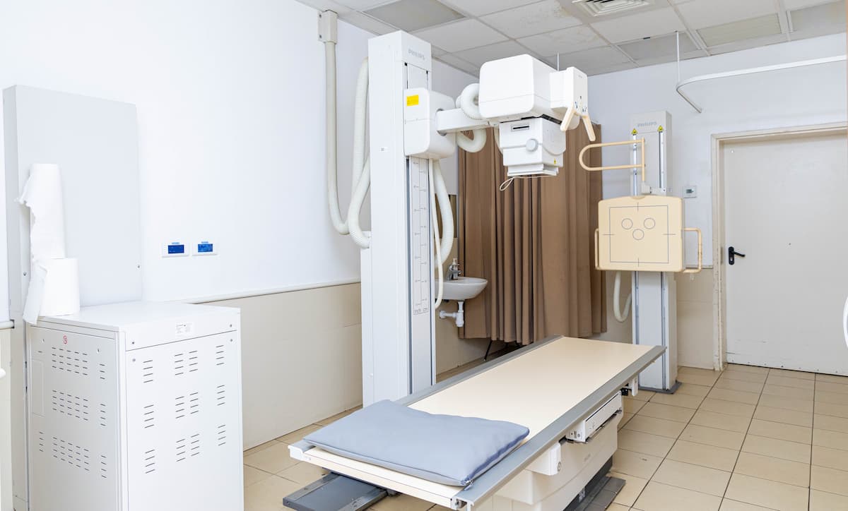
Fluoroscopy
BackFluoroscopy is a specialized imaging service provided by the Radiology Department of the University of Ghana Medical Centre (UGMC). It uses continuous X-ray imaging to produce real-time moving images of internal organs and structures. This allows radiologists to observe bodily functions as they occur and to guide certain diagnostic and interventional procedures with precision.
This modality is commonly used to assess the gastrointestinal tract, urinary system, musculoskeletal system, and reproductive organs, as well as to guide selected diagnostic and interventional procedures.
Fluoroscopy Services By Body Region
1. Gastrointestinal Tract
1. Gastrointestinal Tract
- Barium Swallow, Meal, and Follow-through Studies
- Other Contrast-based Dynamic Studies
2. Urinary Tract
- Micturating Cystourethrogram (MCUG)
- Retrograde Urethrogram (RUG)
- Loopogram
Reproductive / Gynecological
- Hysterosalpingography (HSG)
Fistula & Other Studies
- Fistulogram
Patient Preparation:
Preparation requirements vary depending on the specific fluoroscopic examination:
Preparation requirements vary depending on the specific fluoroscopic examination:
- Gastrointestinal examinations: Patients may be required to fast for several hours before the procedure. In some cases, bowel preparation may be necessary.
- Urogenital studies: Instructions may include arriving with a full or empty bladder, depending on the test.
- Reproductive studies (HSG): Patients are required to bring along a disposable pad and should not have sexual intercourse before the day of the examination.
- General instructions: Patients should wear comfortable clothing and remove metallic items as instructed by personnel. Female patients who are pregnant or suspect pregnancy must inform the radiology personnel prior to the examination.
Note: Patients will receive specific preparation instructions at the time of booking.
Appointment and Time slots:
Fluoroscopy services are provided on week days at 8AM prompt at the Radiology Department. Examinations are performed strictly by appointment. Patients are advised to arrive early to allow adequate time for preparation and registration.
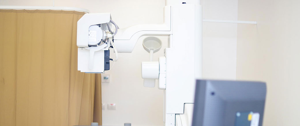
Modality Description:
Fluoroscopy involves the use of a continuous X-ray beam transmitted through the body to a detector, generating live images displayed on a monitor. This allows real – time visualization of physiological processes such as swallowing, digestion, and the movement of contrast material through organs.
Contrast agents including barium or iodine – based media, may be administered orally, rectally, or intravenously to enhance image quality. All procedures are performed by trained radiography staff alongside radiologists, with strict adherence to radiation safety and quality assurance standards.
Other Services
Medical Physics
Medical Imaging
Computed Tomography (CT) Scan
Medical Imaging
Angiography (Cathlab)
Medical Imaging
X-Ray
Medical Imaging
Nanox
Medical Imaging